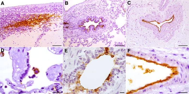
Figure 5.

Immunohistochemistry for Mmm on challenged BREs. Mmm-IR was detectable within the MALT, the IHC pattern resembling that of the dendritic-cells (A; tracheal explant, T24). The presence of Mmm was observed upon the bronchiolar epithelium (B; lung explant, T1), along the endothelial surface of large blood vessel (C; tracheal explant, T24) and inside the cytoplasm of alveolar macrophages (D; lung explant, T1). At higher magnification, the Mmm-IR was apparently seen inside the cytoplasm of bronchiolar epithelial cells (E; lung explant, T1) and of endothelial cells (F; lung explant, T1). Mayer’s hematoxylin counterstain. Scale bar: 10 µm (E), 20 µm (D, F), 50 µm (A), 100 µm (B, C).
