
Fig. 2.
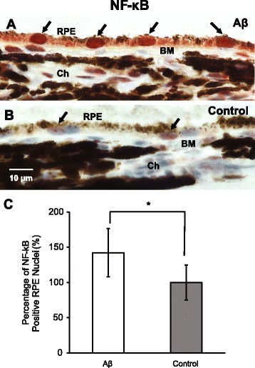
Activation of NF-κB pathway in retinal pigment epithelium (RPE). a–c In retinal cross sections, injections of Aβ enhanced the nuclear translocalization of NF-κB phosphorylated p65 subunit in RPE (garnet red, arrows, a), compared to the light purple RPE nuclei in the control group (arrows, b). By counting the number of RPE nuclei with garnet red (AEC) labeling, there was a ~ 50% increase in the positive RPE nuclei over the control group (c). BM, Bruch’s membrane; Ch, choroid. Scale bar 10 μm. N = 3, Mann-Whitney, *p < 0.05