
Fig. 4.
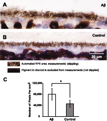
RPE morphological changes following sequential Aβ injections. a Sequential Aβ injections caused RPE swelling, which was quantified by RPE area measurements using a custom Photoshop algorithm identifying the stippled area of RPE pigment. b Animals receiving sequential control solution injections possessed thinner RPE layer compared to the Aβ group, indicated by the stippled area of RPE pigment. c The number of pixels is a surrogate marker for RPE area measurement, which showed about 2-fold increase in Aβ group compared to the control group. N = 3, Mann-Whitney, *p < 0.05