
Fig. 1.
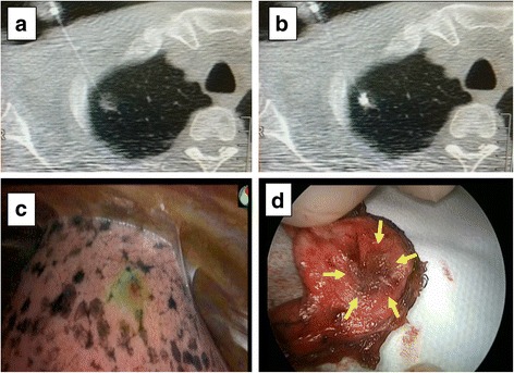
Computed tomography (CT)-guided percutaneous marking, near-infrared (NIR) fluorescence detection, and wedge resection. a: A 23-gauge needle was inserted into the lung near a ground-glass nodule under CT guidance. b: A mixture (0.1 ml) of 100-fold diluted indocyanine green and iopamidol was injected into the lung parenchyma. c: The indocyanine green fluorescence was detected using NIR thoracoscopy (PINPOINT®, Novadaq). d: A resected lung specimen from the surgical field. The target pulmonary nodule is indicated by yellow arrows