Fig. 1.

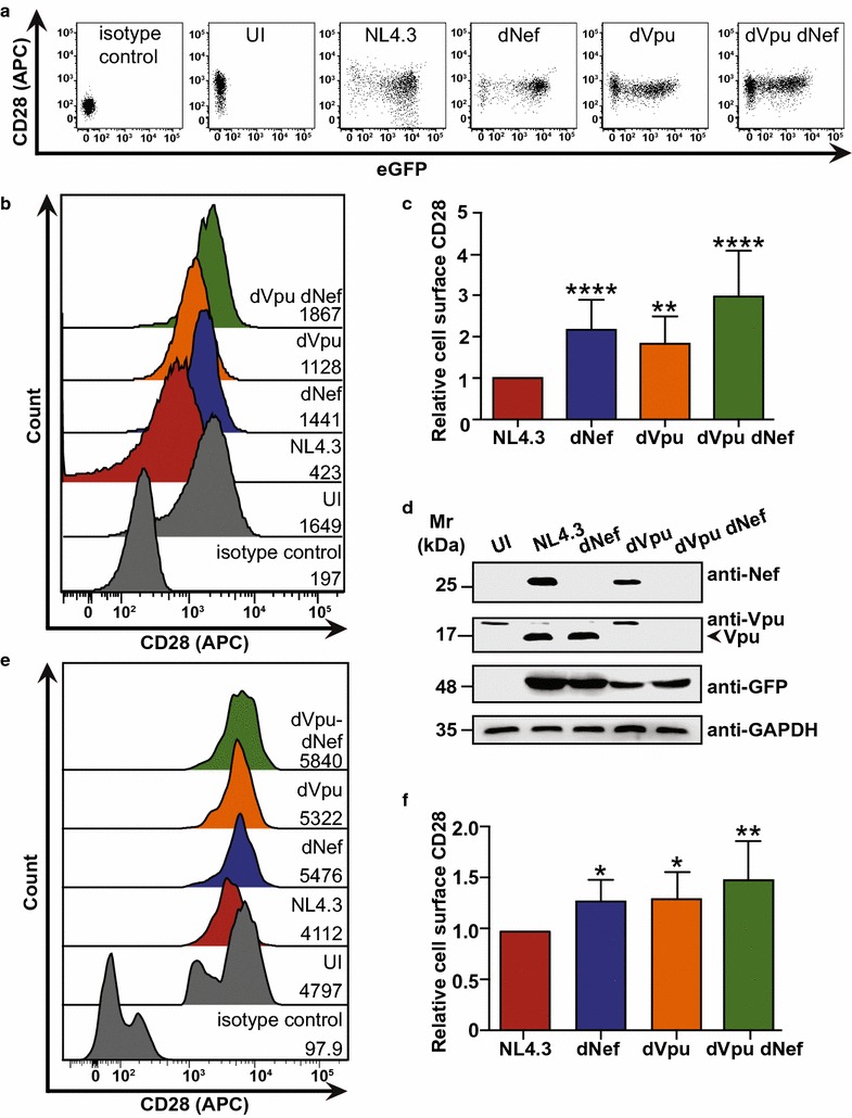
HIV-1 Nef and Vpu downregulate cell surface CD28 protein levels. CD4+ Sup-T1 and primary CD4+ T cells were infected with either VSV-G pseudotyped or replication competent NL4.3, respectively. Viruses encoded Nef and Vpu (NL4.3, red), lacked Nef (dNef, blue) or Vpu (dVpu, orange), or lacked both Nef and Vpu (dVpu dNef, green). Infected cells were stained for CD28 and analyzed by flow cytometry. Live infected Sup-T1 cells were analyzed by gating on Zombie RedTM− and GFP+ cells, and infected primary CD4+ T cells were analyzed by gating on p24+ cells. a Representative dot plots illustrating cell surface CD28 (APC) and infection (GFP+) of live (Zombie RedTM−) Sup-T1 cells. b Representative histograms illustrating cell surface levels of CD28 or the appropriate isotype control on Sup-T1 cells after gating on live (Zombie RedTM−) and infected (GFP+) cells. CD28 (APC) geometric mean fluorescence intensities (MFI) are indicated. c Summary of the relative mean (± SE) cell surface CD28 levels on infected (GFP+) Sup-T1 cells based on MFIs (n = 17). d Western blot illustrating expression of Nef and Vpu in infected Sup-T1 cells. e Representative histograms illustrating cell surface levels of CD28 or the appropriate isotype control on uninfected (UI) or infected (p24+) CD4+ PBMCs. MFIs are indicated. f Summary of the relative mean (± SE) cell surface CD28 levels on infected CD4+ T cells based on MFIs obtained from infection of CD4+ T cells from two healthy donors (n ≥ 3). (UI: uninfected; SE: standard error; Mr: molecular weight; GAPDH: glyceraldehyde 3-phosphate dehydrogenase; GFP: green fluorescent protein; MFI: geometric mean fluorescent intensity; SE: standard error; *p ≤ 0.05; **p ≤ 0.01; ****p ≤ 0.0001)
