
Figure 7. Echocardiographic features in patients receiving extracorporeal support.
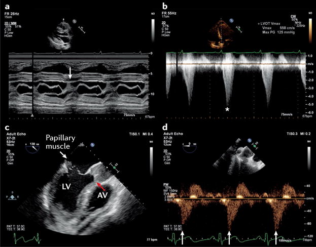
Transthoracic echocardiography in a patient with severe respiratory failure receiving venovenous extracorporeal membrane oxygenation (ECMO). a | Parasternal long axis M-mode echocardiography across the mitral valve showing systolic anterior motion of the mitral valve leaflets (arrow). b | This motion was associated with substantial left ventricular intracavity gradient of 125 mmHg (asterisk). c | A complication of ST-segment elevation myocardial infarction requiring peripheral ECMO is revealed on M-mode echocardiography; papillary muscle rupture had resulted in a flail anterior mitral valve leaflet (white arrow) with associated torrential mitral regurgitation. The increase in left ventricular afterload from ECMO has resulted in failure of the left ventricle (LV) to eject, with a persistently closed aortic valve (AV; red arrow) and stasis of blood in the aortic root. d | Reversal of systolic pulmonary venous flow (arrows) in a patient receiving peripheral venovenous ECMO, suggesting inadequate offloading of the LV.