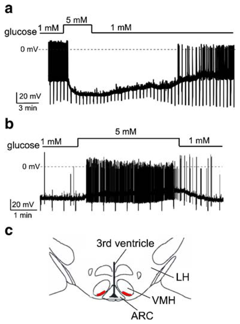
Fig. 1.
Effects of glucose on the membrane potential of VMH neurons dialyzed with high [Cl−]. Current-clamp whole-cell recordings. To monitor membrane resistance, cells were injected with periodic hyperpolarising current pulses (30 pA in a and 20 pA in b). The size of the resulting downward voltage deflections is proportional to membrane resistance. a Representative recording of an inhibitory response. Glucose causes hyperpolarisation, reduces membrane resistance and suppresses firing. b Representative recording of an excitatory response. Glucose causes depolarisation, increases membrane resistance and increases firing. c Region of the VMH where we found glucose-inhibited neurons (red). ARC arcuate nucleus, VMH ventromedial hypothalamus, LH lateral hypothalamus. Slice shown approximately corresponds to adult mouse Bregma coordinates of −1.60 to −1.85 mm

