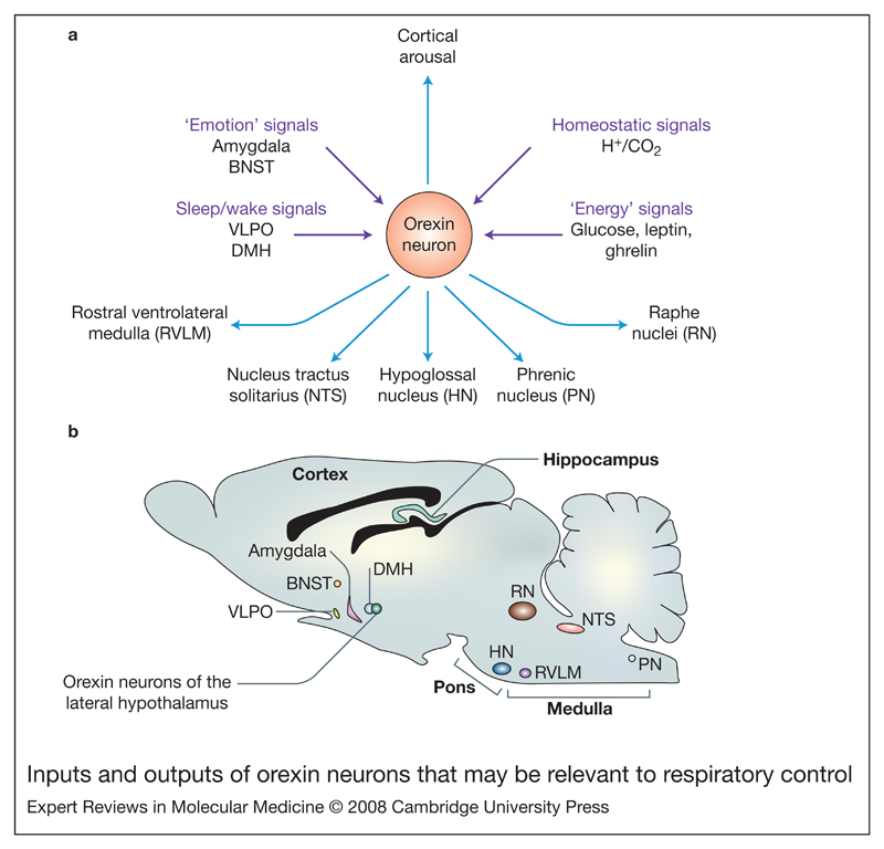
Figure 1. Inputs and outputs of orexin neurons that may be relevant to respiratory control.
(a) Orexin neurons in the lateral hypothalamus send projections both ‘upwards’, to arousal-regulating regions such as the thalamus and the cortex (among other areas – see Ref. 5), and ‘downwards’ to brain stem nuclei directly or indirectly involved in respiratory control: the raphe nuclei, nucleus tractus solitarius (Refs 76, 77), the rostral ventrolateral medulla (Refs 32, 39) and the phrenic and hypoglossal nuclei (Refs 36, 37, 39). In turn, orexin neurons receive anatomical inputs from the amygdala and bed nucleus of stria terminalis (BNST), the dorsomedial hypothalamic nucleus (DMH), GABAergic neurons in the ventrolateral preoptic area (VLPO), and many other hypothalamic and extrahypothalamic areas (see Refs 52, 53). Orexin cells are also able to act as sensors of body energy levels, and acid and CO2 (see Fig. 2). (b) Saggital mouse brain section indicating the location of input and output nuclei mentioned in part a. Abbreviation: GABA, gamma-aminobutyric acid.

