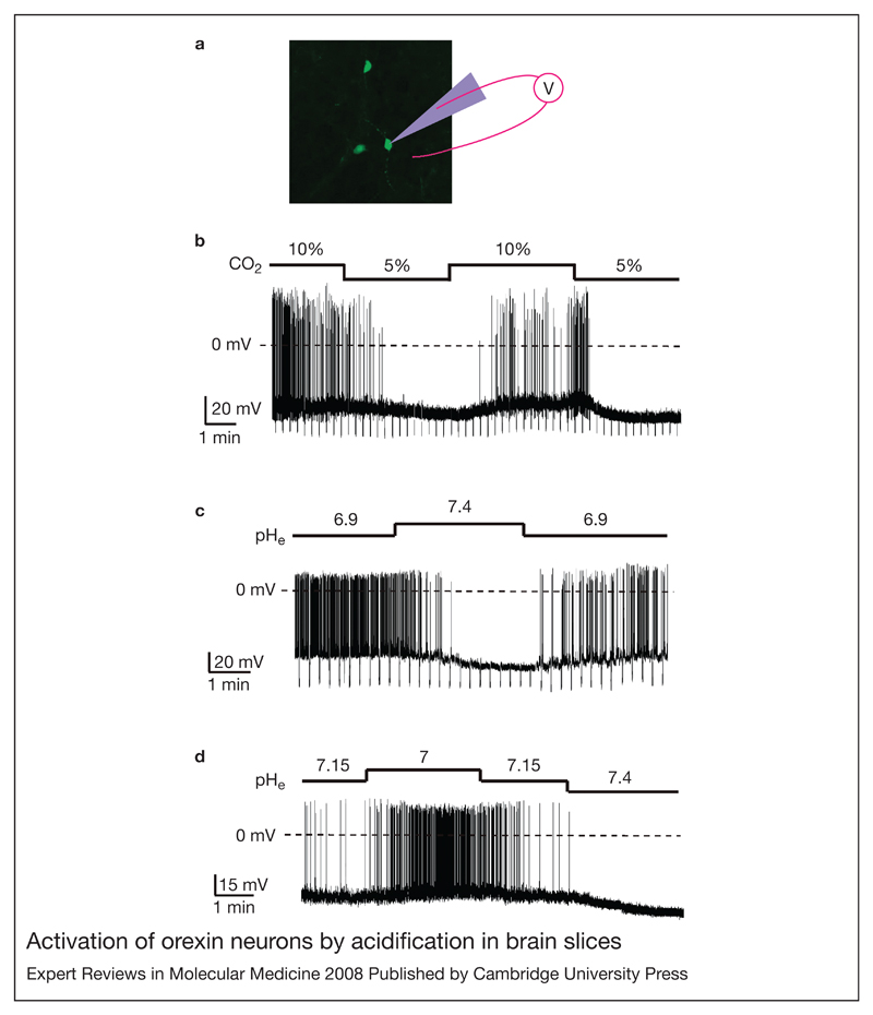
Figure 2. Activation of orexin neurons by acidification in brain slices.
(a) Orexin neurons (green), identified in a mouse brain slice by transgenic green fluorescent protein expression, are shown with a cartoon of a patch-clamp pipette used to measure their membrane potential (full details in Ref. 50). (b) CO2 effects on firing, membrane potential, and membrane resistance of an orexin neuron. (c) pH effects on firing, membrane potential, and membrane resistance of an orexin neuron. (d) Effects of small extracellular pH changes on firing and membrane potential of an orexin neuron. Parts b–d are reprinted from Ref. 50 [Williams, R.H. et al. (2007) Control of hypothalamic orexin neurons by acid and CO2. Proc Natl Acad Sci U S A 104, 10685–10690 (© 2007 by The National Academy of Sciences of the USA)]. Since acid and CO2 levels in the brain are controlled by breathing, modulation of orexin neurons by these signals may communicate breathing state to many brain areas through the widespread projections of orexin cells.

