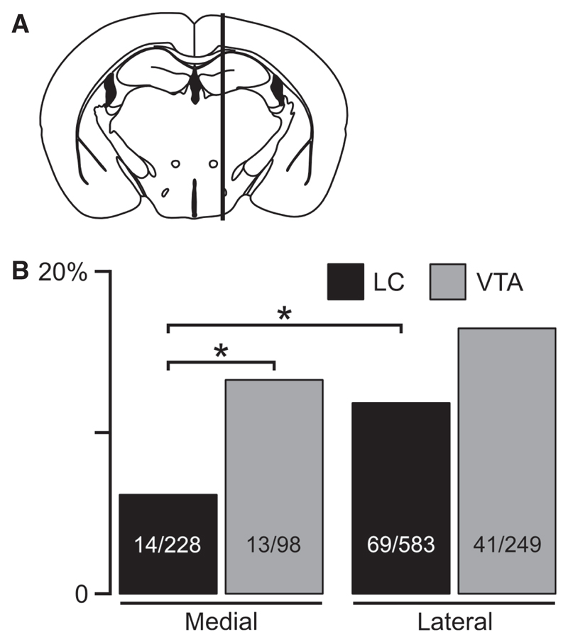
Fig. 3. Medio-lateral distribution of orx/hcrt cells innervating LC or VTA.
(A) The hypothalamus was divided into lateral and medial regions by a vertical line through the fornix (following convention in previous studies, see Results). Orx/hcrt cells that were mapped medial or lateral to this line were grouped together. (B) Proportion of orx/hcrt cells that project to LC or VTA in lateral and medial parts of the lateral hypothalamic area. Only significant differences are indicated (*P < 0.05, Fisher’s exact test). Numbers inside bars are eGFP cells with tracer/total number of eGFP cells.

