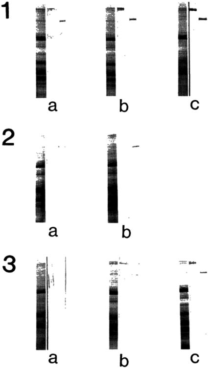
Fig. 1.

Detection of nebulin and connectin in an SDS extract of the biopsied muscle of Duchenne muscular dystrophy. Panel 1, normal; Panel 2, preclinical stage; Panel 3, symptomatic stage. Left lane of each panel, Coomassie Brilliant Blue stained protein bands; Middle lane, immunoblot treated with anti-connectin serum; Right lane
