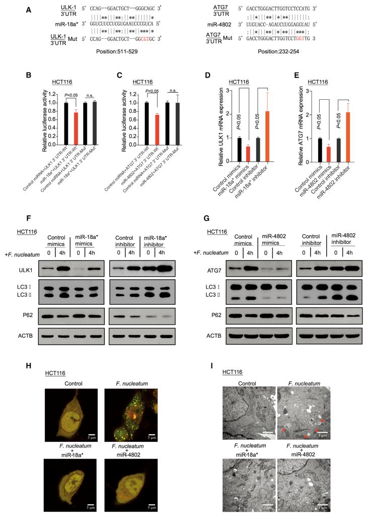
Figure 4. F. nucleatum Activates Cancer Autophagy via Downregulation of miR-18a* and miR-4802.
(A) The predicted binding sequences for miR-18a* (left) and miR-4802 (right) within the human ULK1 and ATG7 3′UTR, respectively. Seed sequences are highlighted.
(B) Luciferase activity was measured in HCT116 cells transfected with miR-18a* mimics or control miRNA. The luciferase reporters expressing wild-type or mutant human ULK1 3′UTRs were used. The luciferase activity was normalized based on the control miRNA transfection. n.s., not significant.
(C) Luciferase activity was measured in HCT116 cells transfected with miR-4802 mimics or control miRNA. The luciferase reporters expressing wild-type or mutant human ATG7 3′UTRs were used.
(D) Real-time PCR was performed in HCT116 cells to detect the expression of ULK1 gene after transfected with miR-18a* mimics or inhibitor, nonparametric Mann–Whitney test.
(E) Real-time PCR was performed in HCT116 cells to detect the expression of ATG7 gene after transfected with miR-4802 mimics or inhibitor, nonparametric Mann–Whitney test.
(F and G) HCT116 cells were transfected with mimics or inhibitor of miR-18a* (F) and miR-4802 (G), respectively. After culturing with F. nucleatum, autophagy and target proteins were detected by western blot in HCT116 cells.
(H) HCT116 cells that stably expressed mRFP-EGFP-LC3 fusion protein were transfected with miR-18a* and miR-4802 mimics. After culturing with F. nucleatum, autophagosomes were observed under confocal microscope (2000 × magnification) in HCT116 cells. Bar scale, 5 μm.
(I) Autophagosomes were observed by transmission electron microscopy (17500 × magnification) in HCT116 cells transfected with miR-18a* (left) and miR-4802 (right) mimics, and then co-cultured with F. nucleatum. Bar scale, 1 μm.
See also Figure S4.

