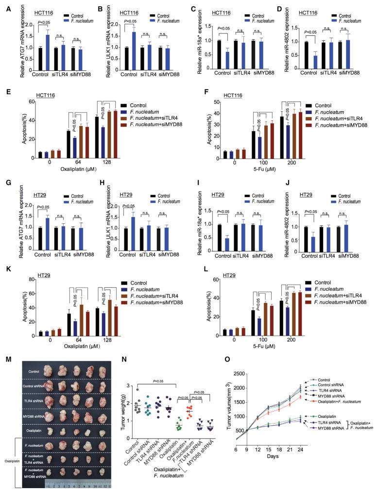
Figure 6. TLR4 and MYD88 Pathway Is Involved in F. nucleatum-Mediated Chemoresistance.
(A–D) Real-time PCR was performed to detect ATG7 (A), ULK1 (B), miR-18a* (C), and miR-4802 (D) expression in HCT116 cells. HCT116 cells were co-cultured with F. nucleatum and transfected with TLR4 and MYD88 siRNAs, respectively.
(E and F) Apoptosis was detected by flow cytometry in HCT116 cells. The cells were co-cultured with F. nucleatum after TLR4 and MYD88 siRNAs transfection, and subsequently treated with different concentrations of Oxaliplatin (E) or 5-FU (F), nonparametric Mann–Whitney test.
(G–J) Real-time PCR was performed to detect expression of ATG7 (G), ULK1 (H), miR-18a* (I), and miR-4802 (J) expression in HT29 cells. HT29 cells were co-cultured with F. nucleatum and transfected with TLR4 and MYD88 siRNAs, respectively.
(K and L) Apoptosis was detected by flow cytometry in HT29 cells. The cells were co-cultured with F. nucleatum after TLR4 and MYD88 siRNAs transfection, and subsequently treated with different concentrations of Oxaliplatin (K) or 5-FU (L), nonparametric Mann–Whitney test.
(M) Representative data of tumors in nude mice bearing HCT116 cells in different groups. Figure 6M and Figure S6A shared experimental controls.
(N and O) Statistical analysis of mouse tumor weights (N) and volumes (O) in different groups, n = 8/group, nonparametric Mann–Whitney test.
See also Figure S7.

