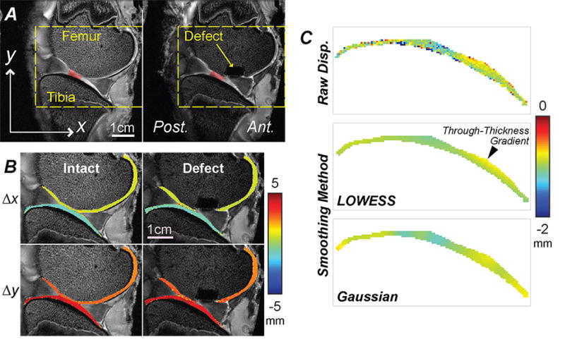
Figure 2. Spatial maps of displacements were noninvasively measured in articular cartilage before and following placement of a critical sized defect in the medial femoral condyle.
Standard MRI of a representative specimen in intact and defect conditions allowed for the identification of a preserved cartilage-cartilage contact region (red shading), and registration of joint morphology in time-sequence MRI scans [A]. In-plane displacements were computed from dualMRI data [B] and show that rigid body motions dominate displacements in the loading direction (y) and direction transverse to loading (x), revealing little obvious internal spatial variations. [C] The raw displacements were smoothed using a locally-weighted linear regression (LOWESS) method, and showed expected through-thickness gradients, in contrast to Gaussian smoothing.

