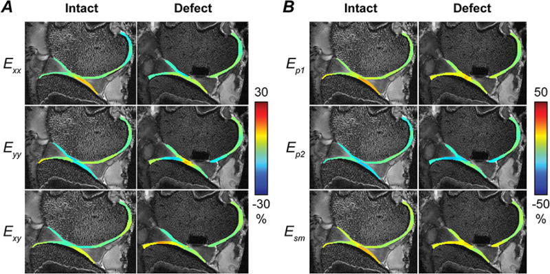
Figure 3. Spatial patterns of strain in articular cartilage increase and localize following creation of a full thickness defect.
In-plane Green-Lagrange strains (Exx, Eyy, Exy) were computed from smoothed displacements [A], and first and second principal (Ep1 and Ep2) and maximum shear (Esm) strains were calculated [B]. High tensile and shear strains were observed at the interface between cartilage-cartilage and cartilage-meniscus contact areas in this representative specimen, with similar high-strain regions observed in other specimens.

