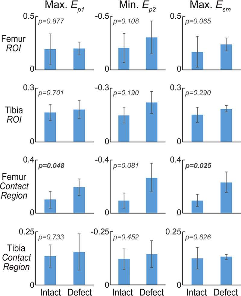
Figure 4. Maximum principal strains in cartilage tibia and femur regions of interest, and contact regions.
Maximum values for first principal and maximum shear strain, and minimum values for second principal strain were computed for full tibial and femoral regions of interest (ROIs) in the intact and defect conditions [A]. The magnitude of principal strains tended to increase with defect placement in femoral, but not tibial, cartilage. Dramatic increases in the maximum shear strain within the contact region of femoral cartilage indicate a heightened sensitivity of this measure to defect placement and altered mechanics within the joint.

