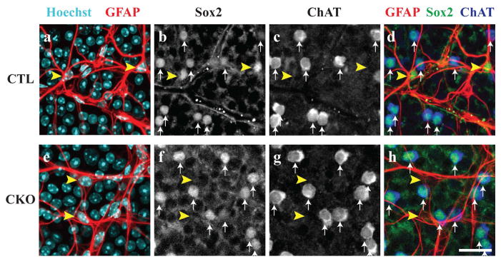
Figure 1. Characterization of Sox2 conditional knockout mice.
In control animals (CTL), astrocytes are visualized by co-localization of Hoechst and GFAP antibodies (yellow arrowheads, a). These cells are distinct from cholinergic amacrine cells that are both ChAT and Sox2-positive (white arrows, b–d). In the Sox2 conditional knockout retinas (CKO), astrocytes are Sox2-deficient as evidenced by the lack of Sox2 staining in astrocyte cell bodies (yellow arrowheads, e–f, h). However, Sox2 remains in cholinergic amacrine cells (white arrows, f–h), showing that Sox2 is removed only from the population of retinal astrocytes. Scale bar = 25 μm.

