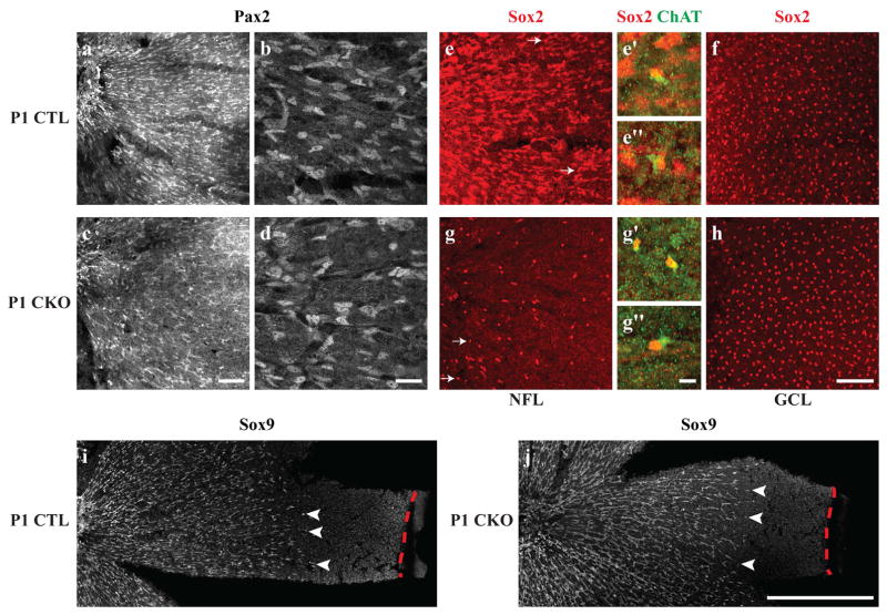
Figure 2. Astrocytes at P1 migrate normally in CKO retinas, despite lacking Sox2.
The migrating wave of astrocytes in both the CTL and CKO retinas, labeled with Pax2, extends from the ONH to about two-thirds of the distance to the retinal margin (a,c). At higher magnification, CTL Pax2-positive astrocytes that are migrating from the ONH in the central retina (left) exhibit an elongated morphology typical of migrating astrocytes (b). CKO Pax2-positive cells appear in comparable density and morphology to CTL retinas (c–d). Almost all Sox2-immunopositive cells in the NFL appear to be astrocytes, with the exception of a few ChAT-positive cells that are occasionally found in the NFL (arrows in e,e′,e″), yet are spatially displaced from the population of Sox2-positive cholinergic amacrine cells in the GCL (f). In CKO retinas, by contrast, very few Sox2-positive cells are found in the NFL (g) and are displaced ChAT-positive cells (g′ and g″). The population of cholinergic amacrine cells in the GCL appears indistinguishable from CTL retinas (h). P1 CTL (i) and CKO (j) retinas were labeled with another astrocytic marker, Sox9, that supports the findings in a–d, that immature astrocytes migrate comparable distances in CTL and CKO retinas (arrowheads indicate the furthest migrating Sox9+ nuclei, while red dashed lines indicate the retinal margin). All panels are oriented with the ONH to the left. Scale bar: a,c,e,f,g,h = 100 μm; b,d = 25 μm; e′,e″,g′,g″ = 10 μm; i,j, = 500 μm.

