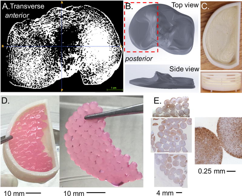
Figure 2.

(A) Transverse slice from high-resolution micro-computed tomography (μCT) of a human cadaveric tibia plateau. (B) SimVascular was used to manually outline the area in each slice and reconstructed to create a steriolithography file (STL), which was imported into SolidWorks as a 3D part. (C) The part was modified to include porous walls and created using a 3D printer. (D) Patient specific METS was created by combining 75 constructs (contact area ~ 950 mm2). (E) Histological staining was positive for aggrecan throughout the construct (brown stain) with cells distributed evenly throughout (blue strain represents cell nuclei).
