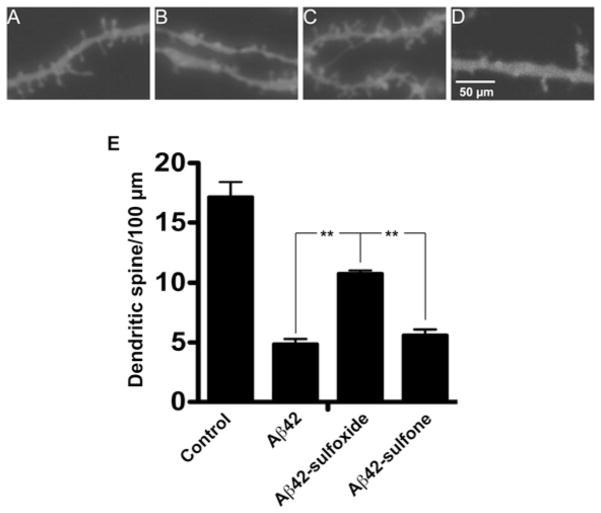
Figure 4. Native and oxidized Aβ42-induced changes in dendritic spine morphology.
Rat primary hippocampal neurons were grown for 21 days, treated with 3 μM of each Aβ42 analogue for 72 h, and stained with DiI. Micrographs are representative of secondary apical branches from multiple neurons. (A) Medium alone, (B) WT Aβ42, (C) Aβ42-sulfoxide and (D) Aβ42-sulfone. (E) The total number of spines in three independent experiments was counted and expressed as means ± S.E.M. **P < 0.01.

