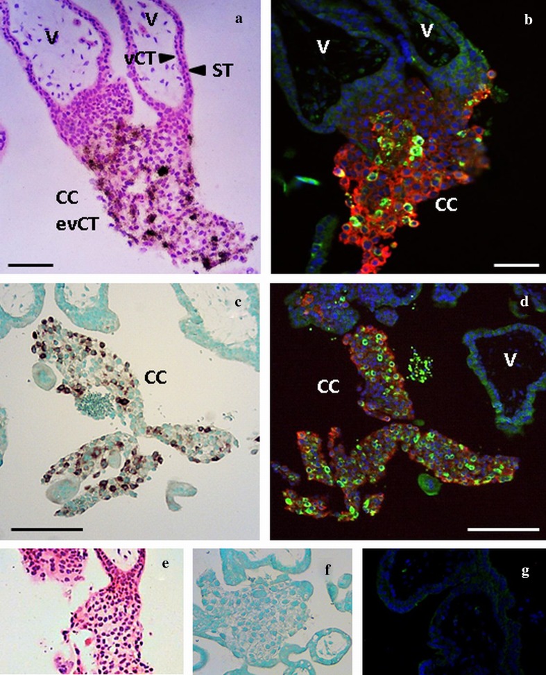
Fig. 2.
Localisation of MMP12 mRNA and protein in the first trimester placenta with radioactive (a) and DIG labelled (c) in situ hybridisation and immunofluorescence (b, d). Cells stained positive for MMP12 mRNA give a black (a) or brown (c) signal in the in situ hybridisation methods. Immunofluorescence costained MMP12 (green) with HLA-G (red). a and b, c and d represents serial sections. a–d MMP12 is located in the trophoblasts of the cell columns (CC) and absent in the placental villi (V). ST syncytiotrophoblast, vCT villous cytotrophoblast, evCT extravillous cytotrophoblasts. Original magnification: ×100. Size of the scale bars: 100 µm (a, b) or 200 µm (c, d). Negative controls for the radioactive (e), DIG labelled (f) and immunofluorescence (g) stainings

