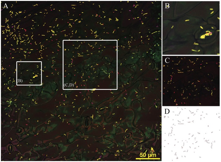
Figure 4.
Composite 3-D image (4 × 4 tiles, approx. 340 μm) showing Sphingomonas, Methylobacterium and Pseudomonas on the leaf surface of in vitro grown Arabidopsis. (A) Hybridization mix included probes EUB338 I, II and III (conjugated with Rhodamine, false colored in red), Methylobacterium mybm-1388 (Alexa-488pink) and Pseudomonas PSE227 (Alexa-647, yellow). (B) Detail showing cells in stomata area, box approximate 50 μm. (C) Image after background removal, box ~100 μm. (D) Result of automatic particle counting, indicating the suitability of images for automatic cell quantification (Fiji). Yellow bar, 50 μm.

