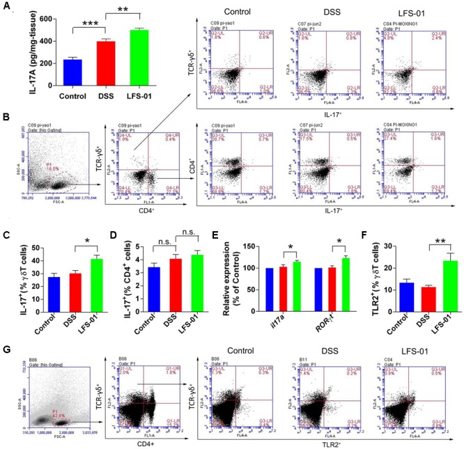
FIGURE 4.

Sulforaphene administration promotesIL-17+ γδT cells in cLP of colitis mice. (A) The IL-17A protein concentration in each animal group was detected in supernatants of colonic tissue homogenates by ELISA. (B) Representative flow cytometry plots of CD4+ T cells identified using side scatter (SSC) and forward scatter (FSC) plots (left panel) and TCR-γδ+expression. Representative flow cytometry plots of Th17 cells (CD4+IL-17+) and γδT17 cells (TCR-γδ+IL-17+)in the LP of mouse colon (right panel). (C). Percentage of γδT17 cells in γδT cells of cLP in mice (n = 7 mice per group). (D) Percentage of Th17 cells in CD4+ T cells of cLP in mice (n = 7). (E) The relative expression levels of il17a and RORγt in colon tissues of mice detected by quantitative real-time PCR (qPCR). (F) Quantification of TLR2 expressing γδT cells (TLR2+TCR-γδ+) in the cLP of mice in different groups. (G) Representative flow cytometry analysis of TLR2 expressing γδT cells. Error bars represent mean ± SEM (n = 7). Statistical analysis of the quantitative multiple group comparisons was performed using the one-way analysis of variance (ANOVA) followed by Tukey’s test, ∗p<0.05, ∗∗p<0.01, ∗∗∗p<0.001; n.s., not significant.
