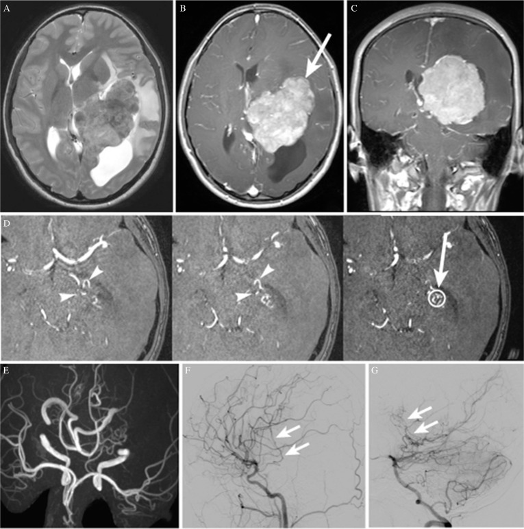
Fig. 1.
(A) Axial T2-weighted magnetic resonance image (MRI) showing a large iso- to hyperintense tumor in the trigone of the left lateral ventricle, associated with peritumoral brain edema and a moderate midline shift to the right. The posterior horn of the lateral ventricle is entrapped. (B, C) Axial and coronal T1-weighted MRI with gadolinium, respectively, showing almost homogeneous tumor enhancement. The white arrow indicates the trajectory followed for tumor resection in the first and the second surgeries. (D) Raw magnetic resonance angiography (MRA) images showing feeders ascending to the antero-inferior surface of the tumor (arrowheads). The open circle indicates the target point for feeder occlusion, and the arrow indicates the trajectory followed to the target. (E) MRA image (axial view) showing the dilated left anterior choroidal and left lateral posterior choroidal arteries. (F) Left carotid angiogram (lateral view) showing feeders originating from the left anterior choroidal artery (arrow). (G) Left vertebral angiogram (lateral view) showing feeders originating from the left lateral posterior choroidal artery (arrow).

