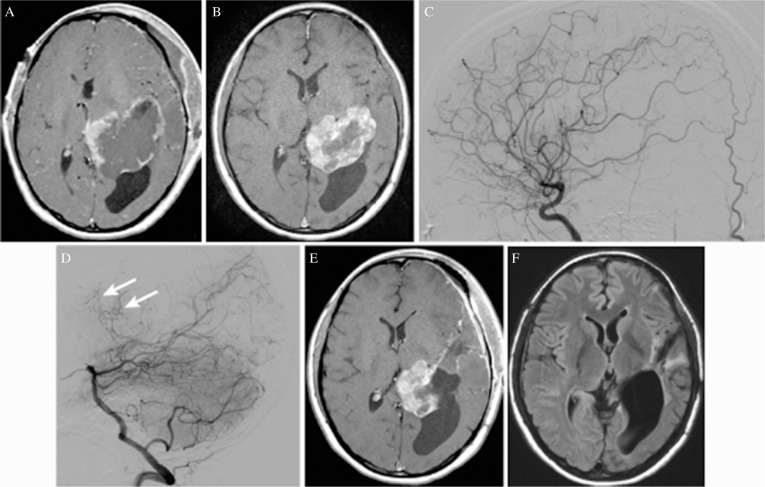
Fig. 3.
(A) Axial T1-weighted magnetic resonance image (MRI) with gadolinium acquired 3 days after the first surgery showing only marginal enhancement of the residual tumor, suggesting that tumor infarction has been induced widely. (B) Axial T1-weighted MRI with gadolinium acquired 1 year after the first surgery showing a remarkable decrease in the volume of the residual tumor (a 20% decrease in diameter) associated with resolution of the midline shift. An increased contrast enhancement area suggests revascularization of the residual tumor. (C) Left carotid angiogram (lateral view) just before the second surgery showing that feeders originating from the left anterior choroidal artery had been obstructed. (D) Left vertebral angiogram (lateral view) just before the second surgery showing remaining or newly developed small feeders originating from the left lateral posterior choroidal artery (arrow). (E) Axial T1-weighted MRI with gadolinium acquired 6 days after the second surgery, showing the remaining medial half of the tumor. (F) Axial fluid-attenuated inversion recovery image acquired 6 days after the third surgery performed by using the high parietal approach, showing complete resection of the tumor.

