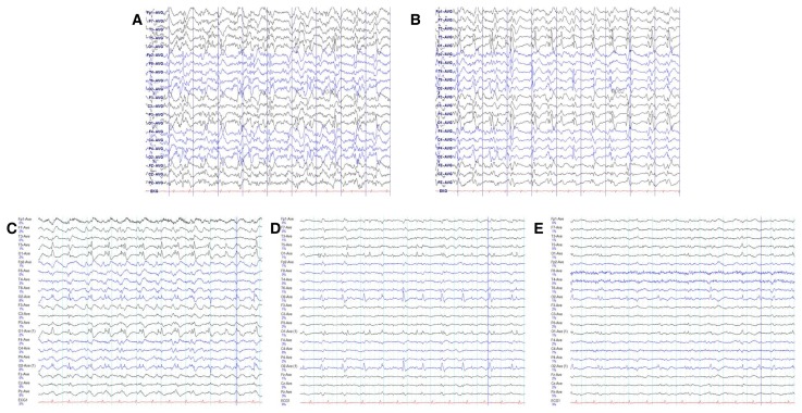
Figure 1.
Ten-second EEG trace recordings with a 10–20 electrode system (A) following initial treatment of convulsive status epilepticus with lorazepam and fosphenytoin loading and showing continuous quasi-periodic single or poly-spikes and waves of high amplitude, mainly in both occipitoparietal areas; (B) after phenobarbital loading and just before induction of TH; (C) following induction of hypothermia to the 35°C target temperature; and after (D) 12 hours and (E) 24 hours of hypothermia at 35°C. EEG, electroencephalography.

