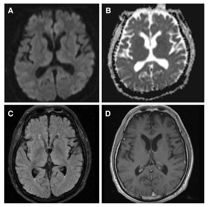
Figure 4.
(A) Brain diffusion, (B) ADC, (C) fluid-attenuated inversion recovery (FLAIR), and (D) T1 enhanced MRI after 2 weeks of TH. Follow-up diffusion MRI and the ADC map showed normalization of the areas with initially reduced diffusion. FLAIR and T1 enhanced MRI showed no abnormal findings. ADC, apparent diffusion coefficient; FLAIR, fluid-attenuated inversion recovery; MRI, magnetic resonance imaging; TH, therapeutic hypothermia.

