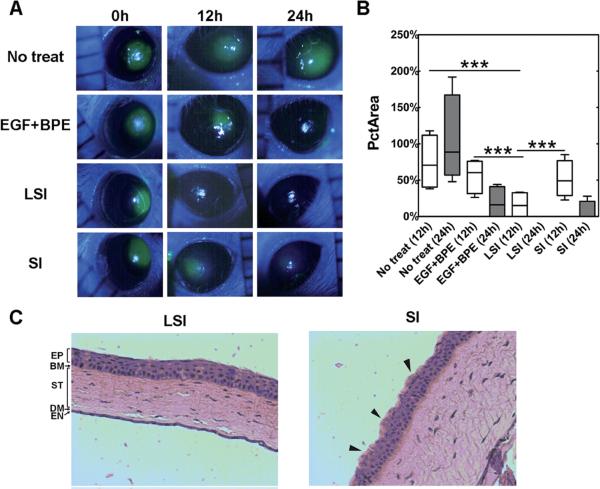
Figure 5. Lacritin-ELP nanoparticles heal corneal wounds in mice.
A 2 mm defect in the corneal epithelium of female non-obese diabetic (NOD) mice was monitored using fluorescein staining at 0, 12 and 24 h with or without treatment by LSI, SI, and a positive control EGF + BPE. (A) Representative images showing the time-lapse healing of the corneal wound. (B) LSI at both 12 and 24 h significantly (***p = 0.001, n = 4) decreased the percentage of initial wound area (PctArea) compared to SI, EGF + BPE, and no treatment groups. (C) After 24 h, corneas were fixed, sectioned across the defect, and stained by hematoxylin and eosin. The corneal epithelium of the LSI treatment group revealed normal pathology. Although reduced fluorescein staining was observed at late times in the SI group, the epithelium did not recover fully, as evidenced by its irregular surface (black arrows). Reprinted from [124] with permission of The Royal Society of Chemistry.

