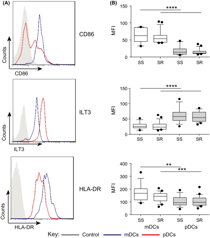
Figure 3.

Expression of surface CD86, ILT3 and HLA‐DR on myeloid (mDCs) and plasmacytoid (pDCs) dendritic cells in the peripheral blood of steroid‐sensitive (SS) and steroid‐resistant (SR) asthmatics. Expression of costimulatory and inhibitory receptors on DCs was assessed by flow cytometric analysis in SS and SR patients at baseline prior to glucocorticoid treatment. A, example histograms of CD86 (top), ILT3 (middle) and HLA‐DR (bottom) expression on mDCs (blue) and pDCs (red) and fluorescence minus one (FMO) control (grey shaded); B, cumulative data on mDCs (white) and pDCs (grey) in the peripheral blood of SS and SR severe asthmatics. Data assessed by paired t test **P < .01, ***P < .001, ****P < .0001
