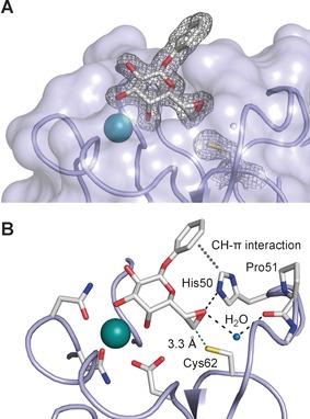
Figure 3.

Crystal structure of epoxide 3 in complex with LecA at 1.80 Å resolution in the non‐covalent binding mode obtained at pH 4.6 (pdb code 5MIH). A) Electron density displayed at 1σ for ligand and Cys62 side chain. B) Interaction of the ligand with LecA: the epoxy oxygen atom accepts hydrogen bonds from His50 and one protein‐bound water molecule. Furthermore, His50 established a CH–π interaction with the phenyl agylycon. In the crystal structure, the sulfur atom of Cys62 is 3.3 Å away from C7 of ligand 3.
