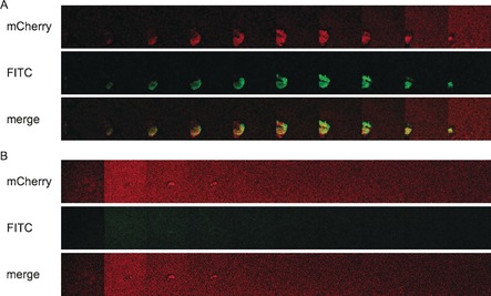
Figure 5.

Galleries of LecA‐dependent staining of P. aeruginosa biofilms with 17. A) P. aeruginosa PAO1 wt or B) the lecA knockout (ΔlecA) mutant, both expressing mCherry from pMP7605, were incubated at 37 °C for 24 h with agitation (180 rpm). Biofilms were stained with the covalent LecA ligand fused to fluorescein (17) for 10–30 min. Z‐stacks (232×232 μm) were recorded every 2 μm at 561 nm for mCherry (red, A and B, upper panels) and 488 nm for fluorescein (green, A and B middle panels). The galleries show every 4th z‐stack recorded. Lower panels show merged images of both channels (488 nm and 561 nm).
