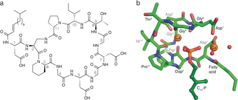
Figure 1.

Laspartomycin C forms a 1:1:2 complex with C10‐P and Ca2+. a) Structure of laspartomycin C. b) Structure of the ternary complex with laspartomycin C (green stick representation), two bound Ca2+ ions (orange spheres), bound water molecules (red spheres), and the C10‐P ligand (dark green ball‐and‐stick representation). The Ca2+ ion on the left is referred to as the central Ca2+ ion and the Ca2+ ion on the right as the peripheral Ca2+ ion.
