
Figure 2.
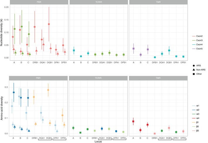
Nucleotide (top) and inferred amino acid (bottom) diversity per site (±σ) at exons encoding the peptide‐binding region (left, with a distinction between antigen‐recognition sites (ARS) and non‐antigen‐recognition sites (non‐ARS) sites); the domains interacting with CD4+ and CD8+ T‐cell receptors (middle); and the trans‐membrane region (right) of the HLA‐A, ‐B, ‐C, ‐DRB1, DQA1, DQB1 and DPB1 molecules in the Mandenka population. Brackets remind the chains forming the HLA‐DQ (DQA1 and DQB1) and HLA‐DP (DPA1 and DPB1) dimers