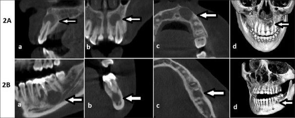
Figure 2.
A: Granuloma in cone beam computed tomography and histology: Patient no. 27. (a) Sagittal view (b) coronal view (c) axial view (d) three-dimensional view. B: Granuloma in cone beam computed tomography and cyst in histology: Patient no. 13. (a) Sagittal view (b) coronal view (c) axial view (d) three-dimensional view

