
Figure 4.
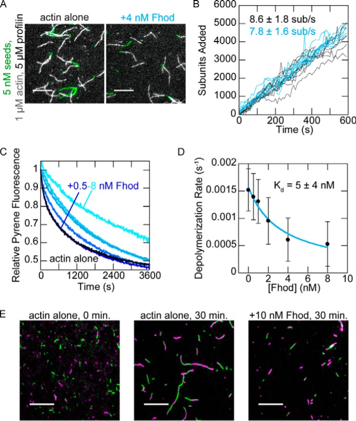
Drosophila Fhod binds barbed ends. A, direct observation of barbed-end elongation by TIRF microscopy with 5 nm F-actin seeds (1% biotinylated, labeled with Alexa Fluor 647-phalloidin), 1 μm G-actin (10% Alexa Fluor 594-labeled), and 5 μm S. pombe profilin, ± 4 nm Fhod. Images were taken 8 min after the start of polymerization. Scale bars, 10 μm. B, quantification of elongation from A. Ten representative traces from each condition are plotted. Elongation rates are average ± standard deviation from three flow chambers for each condition (n = 60, actin alone; n = 36, +4 nm Fhod). C, actin filaments depolymerized by dilution to 0.1 μm (70% pyrene-labeled) with varying concentrations of Fhod. The data were normalized against the initial pyrene fluorescence. D, quantification of depolymerization rates from C, represented by the initial slope over the first 90 s. Fhod binds barbed ends and slows depolymerization. The data and reported Kd are the means ± standard deviation from six independent experiments. The binding curve shows the best fit to the average values. E, reannealing of sheared actin filaments. Actin filaments were stabilized with Alexa Fluor 488- (green) or rhodamine- (magenta) phalloidin, sheared, and allowed to reanneal for 30 min at a concentration of 0.25 μm F-actin ± 10 nm Fhod. Fhod inhibits reannealing of the sheared filaments. Scale bars, 10 μm.