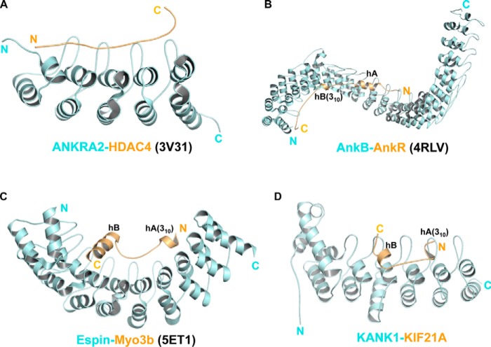
Figure 5.
Comparison of different ankyrin-peptide complex structures. A, the structure of the ANKRA2 ankyrin repeats (cyan) in complex with the HDAC4 peptide (yellow) (PDB code 3V31). B, the complex structure of the AnkB ankyrin domain (cyan) and the AnkR peptide (yellow) (PDB code 4RLV). C, the structure of the Espin ankyrin domain (cyan) with the Myo3b peptide (yellow) (PDB code 5ET1). D, the structure of the KANK1 ankyrin domain (cyan) with the KIF21 peptide (yellow). All ankyrin repeats are arranged from N to C (left to right). The 310-helices of AnkR, Myo3b, and KIF21A are also indicated.

