Figure 1.

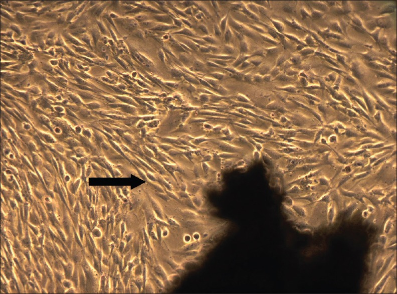
Dental pulp stem cells observed under phase contrast microscope, 7th day (5X magnification)–confluent cell layer observed

Dental pulp stem cells observed under phase contrast microscope, 7th day (5X magnification)–confluent cell layer observed