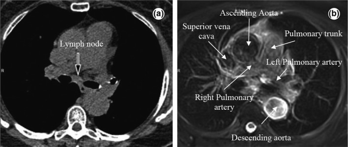
Figure 1.

Axial slices of the mediastinum showing (a) a CT image and the corresponding (b) T2 weighted MRI image with improved soft‐tissue contrast. Some structures (the great vessels) are highlighted in the MRI image. Reprinted from Hochhegger et al.26
