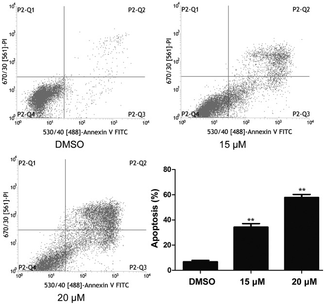
Figure 3.
Lapatinib induced apoptosis of NB4 cells. Cells were treated with 15 and 20 µM lapatinib for 24 h, and apoptosis was analyzed by flow cytometry using double staining with FITC-labeled annexin-V and PI. Cells undergoing early apoptosis are Annexin V-FITC+/PI−, while cells were undergoing late apoptosis are Annexin V-FITC+/PI+. The percentages of late and late apoptotic cells were summed to give the total number of apoptotic cells. Data was expressed as means ± SD. **P<0.01.

