Figure 1.

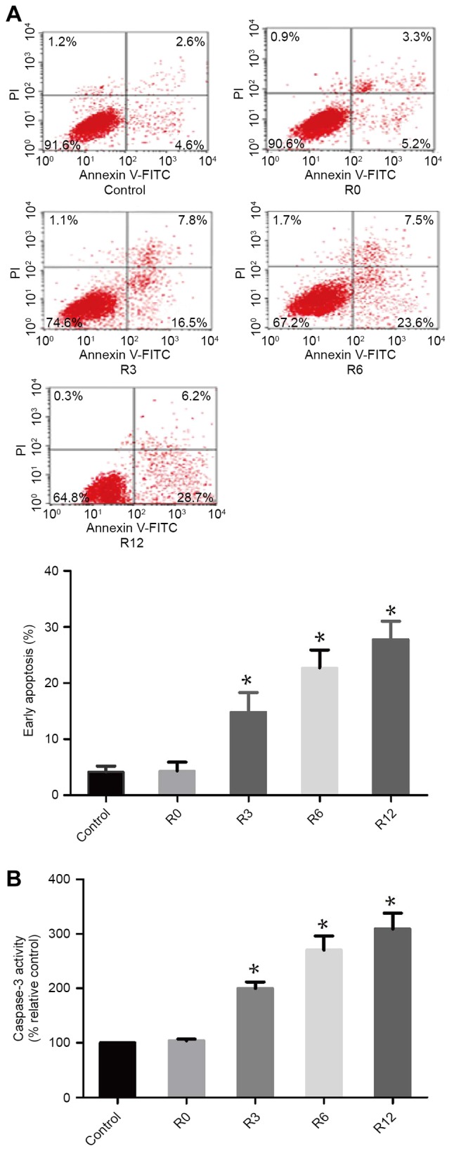
Heat stress induced apoptosis and caspase-3 activation. F98 cells were incubated at 43°C for 60 min to simulate heat stress. Culture medium was then replaced prior to further incubation of the cells for 0, 3, 6 or 12 h (R0 to R12, respectively). (A) Analysis of apoptosis was performed with flow cytometry using Annexin V-FITC/PI staining. (B) The fluorogenic substrate Ac-DEVD-AMC was applied to measure enzymatic activity of caspase-3, which was expressed relative to the control cells incubated at 37°C (set as 100%). *P<0.05 vs. control groups (37°C). FITC, fluorescein isothiocyanate; PI, propidium iodide.
