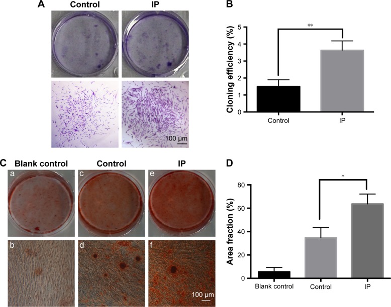
Figure 3.
Effects of IP on colony formation rate (A) and mineralization (C) of hPDLCs. In the mineralization assay, more and larger calcified nodules can be seen in the IP group (e,f) compared with the control group (c,d). And the blank control group (a,b) was used to prove the osteogenic potential of hPDLCs. IP (10−7 M) could stimulate the cloning efficiency (B) and mineralization area fraction (D) of hPDLCs significantly. Mean ± SD (n=3). *P<0.05, **P<0.01 versus control. Magnification ×200.
Abbreviations: IP, ipriflavone; hPDLCs, human periodontal ligament cells; SD, standard deviation.

