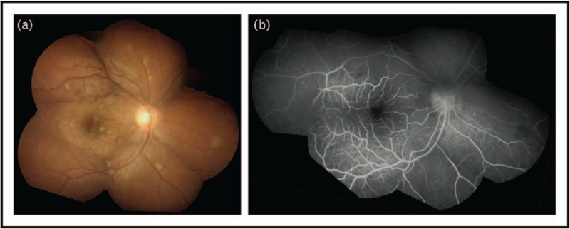
FIGURE 2.

Malaria retinopathy in a Bangladeshi child with cerebral malaria. (A) Composite fundus photograph. (B) Fluorescein angiogram of same fundus image. Images show widespread retinal whitening and patchy hypoperfusion with a white-centered hemorrhage. Typical malarial retinopathy can include four findings: first, macular (perifoveal) and peripheral retinal whitening, second, retinal vessel whitening/discoloration, third, white-centered hemorrhages, and fourth, papilledema. The former two features (first and second) are specific for malaria and the latter two features (third and fourth) are also found in nonmalarial conditions. Reproduced with permission from BMJ Publishing Group Ltd [97].
