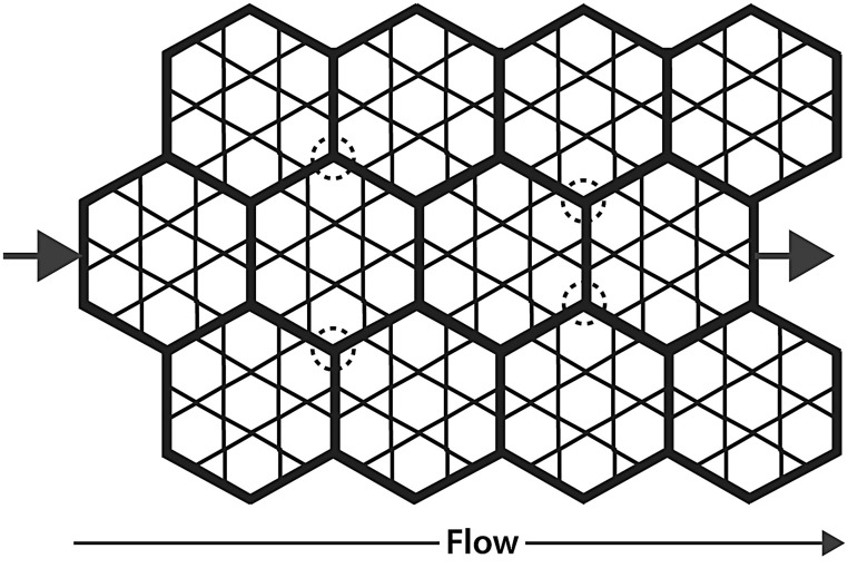
Fig. 6.
Parallel perfusion model of acinar circulation among the alveoli. We hypothesize that the acinar vessels (bold) form a network throughout which perfusion flows among alveoli during flow from arteriole (left arrow) to venule (right arrow). Positive pressure ventilation narrows these vessels, while negative pressure ventilation dilates them. The 4-µm diameter latex particles we infused were confined mainly to these vessels (Fig. 4). Circled areas show examples of network regions that may be less susceptible to compression during positive pressure ventilation, which might be the areas in which particles became trapped in lungs ventilated with positive pressures (Fig. 3c and d).

