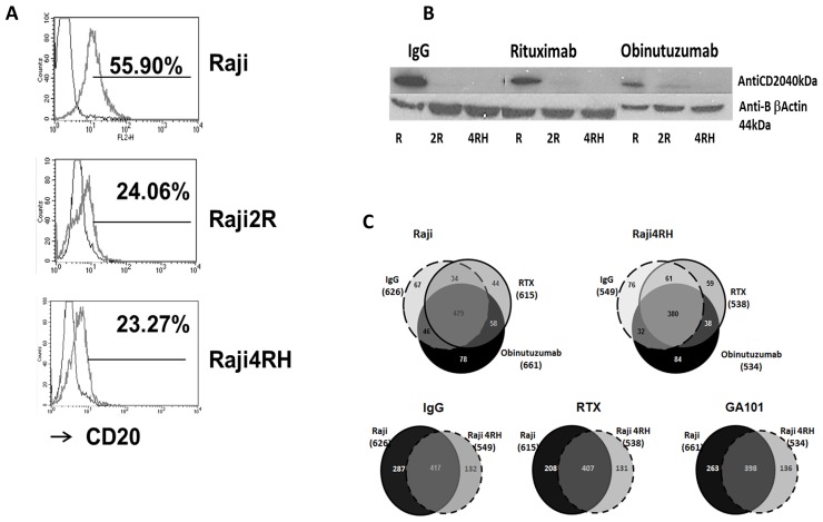
Figure 1. Identification and quantification of phosphorylated proteins in RTX-sensitive and -resistant BL cell lines after obinutuzumab vs. RTX.
(A) CD20 expression: RTX-sensitive (Raji), RTX-resistant (Raji2R and Raji4RH) cell lines were stained with anti-CD20-PE antibody and surface expression of CD20 was determined by flow cytometry (52.3±8.3% vs. 23.9±1.4%, p=0.00003). (B) CD20 expression after obinutuzumab vs. RTX treatment: RTX-sensitive (Raji) and RTX-resistant (Raji2R and Raji4RH) cell lines were treated with 100ug/ml of obinutuzumab (obit), RTX and isotype control IgG for 24 hrs. Cells were lysed and total proteins (40 μg) were suspended in Laemmli sample buffer, boiled, and subjected to SDS polyacrylamide gel electrophoresis on 10% gels for CD20 expression (N=3). (C) Protein identification by phosphoproteomic analysis: Six milligrams of protein from each condition were digested by trypsin and peptides were subjected to phosphopeptide enrichment using metal oxide affinity chromatography (MOAC) and immunoprecipitation for phosphoproteiomics analysis. An LTQ Orbitrap XL in-line with a Paradigm MS2 HPLC was employed for acquiring high-resolution MS and MS/MS data that were searched with the Swissprot Human taxonomic protein database (N=3).

