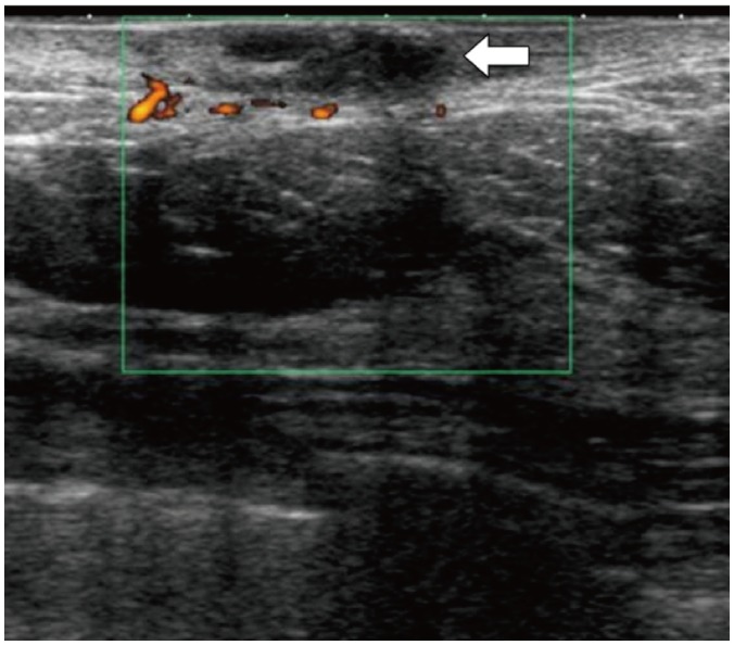
Fig. 9. Infected sebaceous cyst in 59-year-old female with painful left breast lump.

Ultrasound of left breast with color Doppler reveals hypoechoic lesion within dermis with surrounding increased vascularity (white arrow). No deep extension of this lesion is observed. Surrounding skin is thickened.
