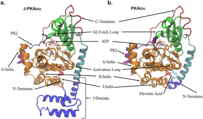
Figure 1.
Structures of J-PKAcα chimera and wild-type PKAcα. (a) J-PKAcα chimera (PDB ID: 4WB7) with the major domains labeled. (b) Wild-type PKAcα (PDB ID: 4DFX). The coloring scheme is as follows: blue = J-domain, Cyan = N-terminal A-helix, Green = Small Lobe, Yellow = Hinge between large and small lobes, Orange = Large Lobe, Red = C-terminal domain, Purple = PKI. ATP, Mg2+ ions, and the myristoylation are shown in licorice representation and colored according to atom.

