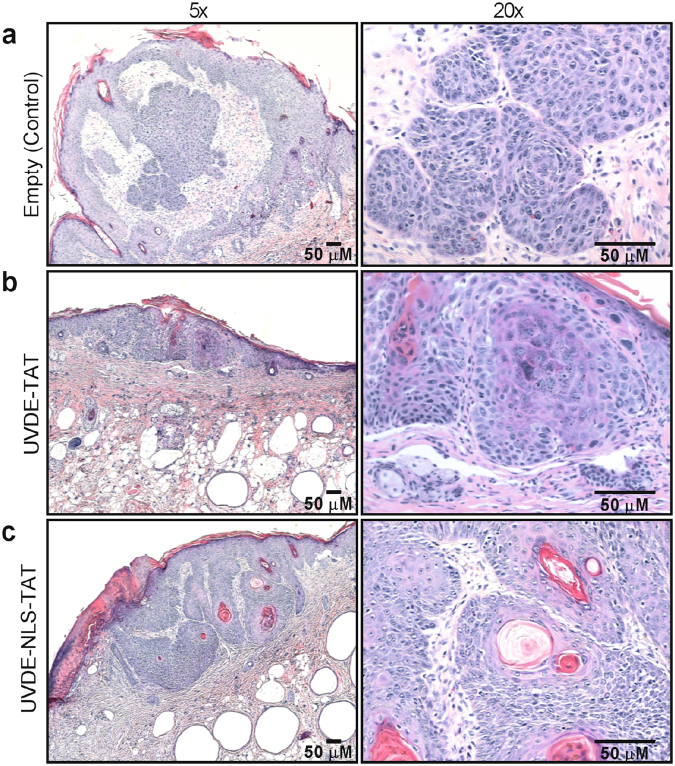
Figure 6.
Representative histology of tumors formed following UVB irradiation. At the time of euthanasia, representative skin tissue samples were harvested, prepared for histologic analyses, and photographed at 5 and 20x in left and right panels, respectively. Representative tumors are shown from skin tissues containing tumors from the following groups: control empty liposomes Panel (a), liposomes containing UVDE-TAT Panel (b), and liposomes containing UVDE-NLS-TAT Panel (c). No qualitative differences were noted in the general characteristics of the tumors that were formed as a result of the cumulative UVB irradiations. All inset bars represent 50 µM.

