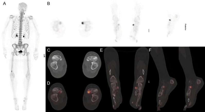
Figure 1.

54-year old female diagnosed with breast cancer presented with left foot pain of 3 months. A. 18F-NaF PET/CT was performed for staging. MIP images demonstrate increased radiotracer uptake in the anteromedial aspect of left mid foot region and right distal femur (bone infarct). B. 18F-NaF PET/CT images show increased radiotracer uptake in the anteromedial aspect of left mid foot. C. Axial CT images demonstrate a bilateral osseous density medial to the navicular bone which was suggestive of a Type II accessory navicular bone. D-F. Fused 18F-NaF PET/CT shows increased tracer uptake in the articulation with sclerosis between the medial border of the left navicular and an os naviculare. Scan appearances are suggestive of left painful accessory os syndrome.
