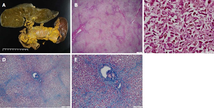
Figure 4.

Pathological findings in the liver. A: The liver (1580 g) was bile stained and soft; B: Low power field: The capsule was wrinkled in HE staining; C: High power field: Microscopically, the liver showed massive necrosis and collapse of the intervening parenchyma in HE staining; D: Low power field in AZAN staining; E: High power field in AZAN staining: Bridging pattern of fibrosis suggestive of chronic liver injury was not found. HE: Hematoxylin-eosin.
