Figure 3.

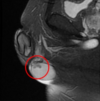
Magnetic resonance imaging‐realized T2‐weighted, low‐sign, bilateral, solid enlarged serpiginous lesions in the testicular parenchyma

Magnetic resonance imaging‐realized T2‐weighted, low‐sign, bilateral, solid enlarged serpiginous lesions in the testicular parenchyma